Home » Without Label » Back Muscles Anatomy - Anatomy of human superfacial muscles back vintage ... - Muscles of the back can be divided into superficial, intermediate, and deep group.
Back Muscles Anatomy - Anatomy of human superfacial muscles back vintage ... - Muscles of the back can be divided into superficial, intermediate, and deep group.
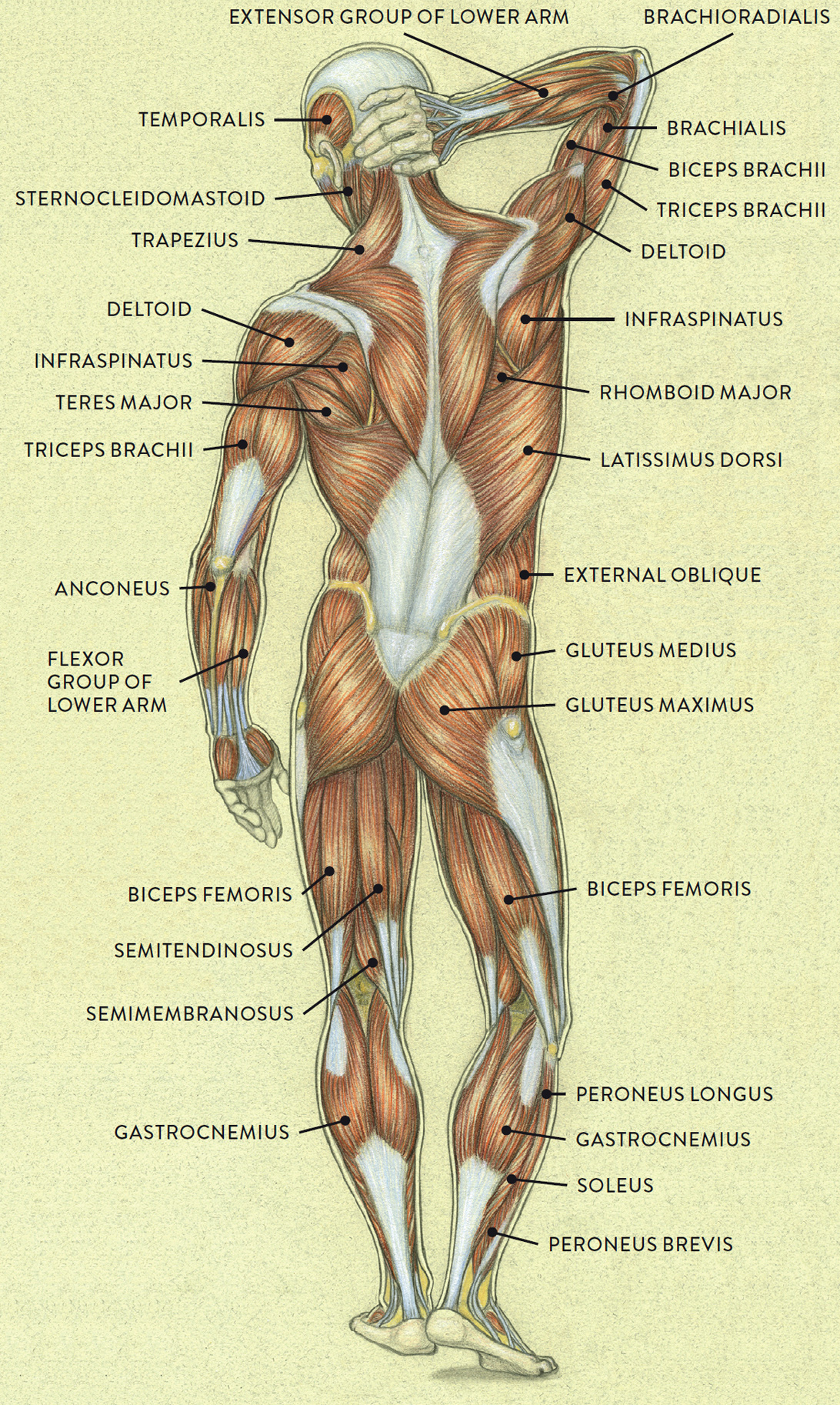
Back Muscles Anatomy - Anatomy of human superfacial muscles back vintage ... - Muscles of the back can be divided into superficial, intermediate, and deep group.. Anterior rami of spinal nerve innervate them. Balance the weight of your head on top of your spine evenly distribute weights from your upper body into the lower extremities A very important muscle in terms of body beauty is responsible for giving the shape of the v that indicates a macho back. This curve, called lordosis, helps to: The superficial back muscles are situated underneath the skin and superficial fascia.
The tendon for latissimus dorsi can be harvested for tendon repairs or tendon transfers. Back muscles the muscles of the back are a group of strong, paired muscles that lie on the posterior aspect of the trunk. Muscle or ligament strains can occur from repeated use of the muscles, or from improperly or awkwardly lifting heavy objects. They start at the top of the neck and go down to the tailbone. Anatomynote.com found anatomy of back muscles diagram from plenty of anatomical pictures on the internet.
MUSCLES OF THE FEMALE FIGURE—ANTERIOR VIEW from schoolbag.info Balance the weight of your head on top of your spine evenly distribute weights from your upper body into the lower extremities The deltoid, teres major, teres minor, infraspinatus, supraspinatus (not shown) and subscapularis muscles (not shown) all extend from the scapula to the humerus and act on the shoulder joint. Browse 3,575 back muscle anatomy stock photos and images available, or search for pelvic bone or lymphatic system to find more great stock photos and pictures. The back consists of the spine, spinal cord, muscles, ligaments, and nerves. A very important muscle in terms of body beauty is responsible for giving the shape of the v that indicates a macho back. Latissimus dorsi is a large muscle that, when atrophied, can cause significance asymmetry in the back. The back comprises interconnecting nerves, bones, muscles, ligaments, and tendons, all of which can be a source of pain. The upper back is a complex area containing a number of muscles that perform various actions on the scapulae (shoulder blades) and humerus.
The abdominal muscles also play a major role in the posture and stability to the body and compress the organs of the abdominal cavity during various activities such as breathing and defecation.
On this page, you'll learn about each of these muscles, their locations and functional anatomy. They provide movements of the spine , stability to the trunk, as well as the coordination between the movements of the limbs and trunk. Your lower back (lumbar spine) is the anatomic region between your lowest rib and the upper part of the buttock. The back muscles are anatomically layered into superficial (extrinsic) and deep (intrinsic) muscles. Anatomy of the upper back. The abdominal muscles also play a major role in the posture and stability to the body and compress the organs of the abdominal cavity during various activities such as breathing and defecation. Lower back muscles anatomy 12 photos of the lower back muscles anatomy lower back muscle energy techniques, lower back muscles damage, lower back muscles pulsating, lower back muscles sore, lower back muscles tighten up when walking, human muscles, lower back muscle energy techniques, lower back muscles damage. Since the all the back muscles originate in embryo (fetus) form by locations other than the back, muscles in the superficial, as well as, intermediate groups, are extrinsic muscles. Anatomy of back muscles your back consists of three distinct layers of muscles, namely the superficial layer, the intermediate layer, and the deep layer. While it is a strong muscle for adduction, internal rotation and extension of the humerus, we can do without it. What are the lower back muscles and their anatomy? The deep muscles develop in the back called intrinsic muscles. All about the back muscles the back anatomy includes the latissimus dorsi, trapezius, erector spinae, rhomboid, and the teres major.
Back muscles the muscles of the back are a group of strong, paired muscles that lie on the posterior aspect of the trunk. By far the most common cause of back pain is muscle strain. The spaces between the vertebrae are maintained by intervertebral discs that act like shock absorbers throughout the spinal column to cushion the bones as the body moves. The upper back is a complex area containing a number of muscles that perform various actions on the scapulae (shoulder blades) and humerus. Latissimus dorsi is a large muscle that, when atrophied, can cause significance asymmetry in the back.
Drawsh: Sacro Spinalis from 2.bp.blogspot.com Understanding lower back anatomy is key to understanding the root of lower back and hip pain. The trapezius and latissimus dorsi muscles connect the upper limb to the vertebral column. They start at the top of the neck and go down to the tailbone. 1 your spine in this region has a natural inward curve. Muscle or ligament strains can occur from repeated use of the muscles, or from improperly or awkwardly lifting heavy objects. Anatomy of the back muscles the latissimus dorsi muscles (also known as the lats) are the largest muscles of the back. The spaces between the vertebrae are maintained by intervertebral discs that act like shock absorbers throughout the spinal column to cushion the bones as the body moves. We think this is the most useful anatomy picture that you need.
Anatomynote.com found anatomy of back muscles diagram from plenty of anatomical pictures on the internet.
All about the back muscles the back anatomy includes the latissimus dorsi, trapezius, erector spinae, rhomboid, and the teres major. The back comprises interconnecting nerves, bones, muscles, ligaments, and tendons, all of which can be a source of pain. The superficial back muscles are situated underneath the skin and superficial fascia. Understanding lower back anatomy is key to understanding the root of lower back and hip pain. Anatomynote.com found anatomy of back muscles diagram from plenty of anatomical pictures on the internet. These muscles include the large paired muscles in the lower back, called erector spinae, which help hold up the spine, and gluteal muscles. The deltoid, teres major, teres minor, infraspinatus, supraspinatus (not shown) and subscapularis muscles (not shown) all extend from the scapula to the humerus and act on the shoulder joint. These sections are cervical (neck), thoracic (upper and middle back), lumbar (lower back), and sacrum (tailbone). Back pain is the second most common type of pain in adults (the most common being headaches). We think this is the most useful anatomy picture that you need. The muscles of the lower back, including the erector spinae and quadratus lumborum muscles, contract to extend and laterally bend the vertebral column. Browse 3,575 back muscle anatomy stock photos and images available, or search for pelvic bone or lymphatic system to find more great stock photos and pictures. Superficial back muscles, intermediate back muscles and intrinsic back muscles.the intrinsic muscles are named as such because their embryological development begins in the back, oppose to the superficial and intermediate back muscles which develop elsewhere and are therefore classed as extrinsic muscles.
The lower back (where most back pain occurs) includes the five vertebrae in the lumbar region and supports much of the weight of the upper body. These muscles give height and breadth to back development. Back muscles anatomy the surface muscles of the upper back include the trapezius muscles (traps) and posterior deltoids. The tendon for latissimus dorsi can be harvested for tendon repairs or tendon transfers. Anatomynote.com found anatomy of back muscles diagram from plenty of anatomical pictures on the internet.
Anatomy of human superfacial muscles back vintage ... from i.pinimg.com The extensor muscles are attached to back of the spine and enable standing and lifting objects. Back pain is one of the most common kinds of pain for adults, and muscle strains are the most common type of back pain. Muscles of the lumbar spine. Anatomy of back muscles your back consists of three distinct layers of muscles, namely the superficial layer, the intermediate layer, and the deep layer. The back muscles are anatomically layered into superficial (extrinsic) and deep (intrinsic) muscles. The spaces between the vertebrae are maintained by intervertebral discs that act like shock absorbers throughout the spinal column to cushion the bones as the body moves. They provide movements of the spine , stability to the trunk, as well as the coordination between the movements of the limbs and trunk. Anatomy of the back muscles the latissimus dorsi muscles (also known as the lats) are the largest muscles of the back.
The human spine is composed of 4 sections of vertebrae.
The back comprises interconnecting nerves, bones, muscles, ligaments, and tendons, all of which can be a source of pain. All these muscles are therefore associated with movements of the upper limb. Browse 3,575 back muscle anatomy stock photos and images available, or search for pelvic bone or lymphatic system to find more great stock photos and pictures. The upper back is a complex area containing a number of muscles that perform various actions on the scapulae (shoulder blades) and humerus. Anterior rami of spinal nerve innervate them. The back muscles are anatomically layered into superficial (extrinsic) and deep (intrinsic) muscles. The superficial back muscles are situated underneath the skin and superficial fascia. This curve, called lordosis, helps to: Lower back muscles anatomy 12 photos of the lower back muscles anatomy lower back muscle energy techniques, lower back muscles damage, lower back muscles pulsating, lower back muscles sore, lower back muscles tighten up when walking, human muscles, lower back muscle energy techniques, lower back muscles damage. On this page, you'll learn about each of these muscles, their locations and functional anatomy. Muscles of the back can be divided into superficial, intermediate, and deep group. We think this is the most useful anatomy picture that you need. A very important muscle in terms of body beauty is responsible for giving the shape of the v that indicates a macho back.